
Interventional Radiologist & Endovascular Surgeon — Trained at Mount Sinai & Columbia University / NYP, New York. Founder of Israel Vein Center and Senior IR at Shamir Medical Center.
Dr. Shimon Aronhime, MD — Interventional Radiologist & Endovascular Surgeon
📍 Lilienblum 17, Tel Aviv | Shamir Medical Center, Beer Yaakov
📞 073-374-4040 |
You’ve had a duplex Doppler ultrasound for varicose veins and now have a report in hand. The medical terminology can seem impenetrable. This guide explains every major finding you’re likely to encounter.
How to Read Your Venous Doppler Ultrasound Report — Patient Guide
What Is a Venous Doppler Ultrasound?
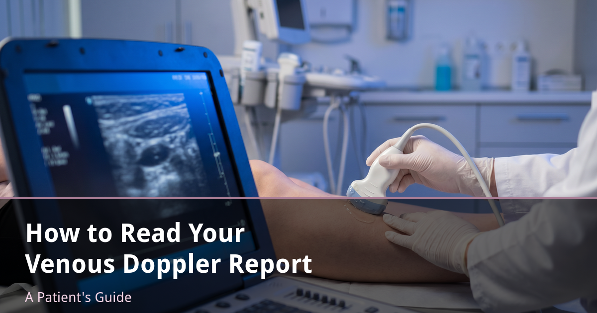
A venous duplex ultrasound is a real-time imaging study that combines B-mode (grayscale anatomical imaging) with colour Doppler (blood flow direction and velocity). It is the gold standard for diagnosing chronic venous insufficiency, mapping reflux, and planning treatment.
The study is performed with you standing (or tilted upright) so gravity maximises the demonstration of reflux. The sonographer compresses the calf or thigh and watches whether blood flows backward when released — a finding called reflux.
CEAP Classification — What Your Grade Means
CEAP is the international classification system for chronic venous disease. The “C” (Clinical) component runs from C0 to C6:
- C0 — No visible or palpable signs of venous disease
- C1 — Telangiectasias or reticular veins (spider veins, small blue veins)
- C2 — Varicose veins (>3mm diameter)
- C3 — Oedema (swelling) without skin changes
- C4a — Pigmentation or eczema
- C4b — Lipodermatosclerosis or atrophie blanche
- C5 — Healed venous ulcer
- C6 — Active venous ulcer
C1–C2 are usually cosmetic or mildly symptomatic. C3–C6 indicate progressive venous insufficiency that typically requires medical treatment.
Reflux — Duration and Significance
Reflux is measured in seconds. Normal venous closure time after calf compression is <0.5 seconds. Pathological reflux is defined as:
- Great saphenous vein (GSV): reflux >0.5 seconds
- Small saphenous vein (SSV): reflux >0.5 seconds
- Deep veins: reflux >1.0 second
The duration of reflux tells you the severity of valve failure. Reflux lasting >2–3 seconds typically indicates significant insufficiency requiring treatment.
Key Abbreviations in Your Report
- GSV — Great saphenous vein (long vein running up the inner leg)
- SSV — Small saphenous vein (behind the knee)
- SFJ — Saphenofemoral junction (where the GSV meets the femoral vein at the groin)
- SPJ — Saphenopopliteal junction (where the SSV meets the popliteal vein behind the knee)
- CFV — Common femoral vein
- Pop V — Popliteal vein
- PR — Proximal reflux / perforator reflux
- DVT neg — No deep vein thrombosis found
Vein Diameter — What Numbers Mean
- GSV <5mm — Normal; sclerotherapy may be suitable for symptomatic cases
- GSV 5–10mm — Thermal ablation (EVLA or RFA) is appropriate
- GSV >10mm — VenaSeal is particularly effective for larger, tortuous veins; thermal ablation also possible
What “Incompetent SFJ” Means
The saphenofemoral junction (SFJ) is where the great saphenous vein joins the deep venous system at the groin. “Incompetent SFJ” means the valve at this junction is failing, allowing blood to flow backward down the entire length of the GSV. This is the most common finding in truncal venous insufficiency and typically requires thermal ablation or VenaSeal of the GSV.
Treatment Implications
Here’s how findings typically translate to treatment:
- Incompetent SFJ + GSV reflux → EVLA, RFA, or VenaSeal of GSV
- Isolated SSV reflux → EVLA or RFA of SSV
- Residual tributaries after truncal treatment → sclerotherapy
- C1 spider veins only → cosmetic sclerotherapy
- Incompetent perforators → ultrasound-guided foam sclerotherapy
Further Reading
Ready to discuss your treatment options?
Ready to Book a Consultation?
Duplex ultrasound mapping included at the first visit
Israel Vein Center — Lilienblum 17, Tel Aviv