
 Dr Shimon Aronhime MD/venous-doppler-ultrasound-israel.png)
Venous Doppler Ultrasound — What Patients Need to Know
Endovascular Surgeon & Interventional Radiologist — trained at Mount Sinai & Columbia University / NYP, New York. Founder of Israel Vein Center; Senior Attending, Shamir Medical Center.
What Is Venous Duplex Ultrasound?
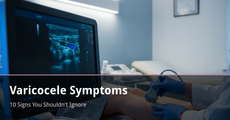
A venous duplex ultrasound — also called venous duplex scan or Doppler mapping — combines two imaging modalities in a single examination. B-mode ultrasound creates a real-time anatomical image of the venous structures: their diameter, wall integrity, and course. Doppler technology measures the speed and direction of blood flow within those structures. Together, they provide the complete picture of venous function that no physical examination alone can supply.
The procedure uses no radiation, requires no injections or contrast, causes no pain, and takes approximately 20–30 minutes.
What the Examination Reveals
- Venous reflux — backward (downward) blood flow indicating valve incompetence, the primary cause of varicose veins
- Vein diameter — great saphenous vein (GSV) diameter above 6–7 mm is considered dilated
- Valve function — whether valves close adequately on standing and with Valsalva maneuver
- Deep vein thrombosis (DVT) — critical safety information before any superficial vein procedure
- Perforator incompetence — abnormal connections between superficial and deep systems
- Anatomical map — the course of GSV, SSV, and their junctions — the roadmap for treatment planning
Understanding Venous Reflux
In healthy leg veins, blood flows upward toward the heart. Venous valves — small bicuspid structures spaced throughout the superficial and deep veins — act as one-way gates, preventing reversal. When valves fail (through genetics, pregnancy, prolonged standing, obesity, or prior thrombosis), blood falls back and accumulates in the veins below.
During duplex examination, the Valsalva maneuver or manual compression-release provokes this reversal. Duration of retrograde flow is measured. Reflux exceeding 0.5 seconds in the great saphenous vein (GSV) or 0.35 seconds in the small saphenous vein (SSV) is pathological. This single measurement determines whether treatment is warranted and what type.
CEAP Classification — From Scan to Diagnosis
Duplex findings, combined with clinical examination, allow CEAP classification of chronic venous disease:
- C0/C1 — no visible disease / spider veins only
- C2 — varicose veins ≥ 3 mm
- C3 — venous edema (leg swelling)
- C4 — skin changes: hyperpigmentation, eczema, lipodermatosclerosis
- C5/C6 — healed or active venous ulcer
CEAP classification determines treatment urgency, guides insurance coverage decisions, and forms the basis of the medical record.
Why Duplex Is Mandatory Before Treatment
Visible varicose veins are rarely the problem — they are a symptom. The source is typically a refluxing trunk vein higher in the leg, not visible to the naked eye. Treating only what you can see while missing the upstream source results in early recurrence. The duplex map tells the clinician exactly what to treat, where to start, and in what order.
Duplex at Israel Vein Center
Dr. Aronhime performs the examination himself, at the same visit as your consultation. There is no separate radiology referral, no waiting weeks for a report, and no risk of information being lost between a radiologist and a treating physician. The scan is performed in both supine and standing positions — essential for detecting postural reflux that may be absent lying down. Findings are immediately integrated into the treatment plan.
Book a consultation with duplex included: Contact us | +972-73-374-4040
Frequently Asked Questions
What does a venous duplex ultrasound show?
Venous duplex combines B-mode ultrasound imaging with Doppler flow measurement. It shows: venous reflux (backward flow indicating valve failure), vein diameter and wall integrity, deep vein thrombosis, perforator incompetence, and the anatomical map of the superficial venous system needed to plan treatment. No radiation, no pain, approximately 20–30 minutes.
Is duplex ultrasound required before every varicose vein treatment?
Yes, without exception. Treating varicose veins without prior duplex mapping is like operating without imaging. The duplex identifies which trunk vein is responsible, where reflux originates, and which tributaries it supplies — determining exactly what to treat and in what order. Without mapping, you risk treating downstream tributaries while missing the source, leading to early recurrence.
What does “venous reflux” mean?
Reflux means blood flowing in the wrong direction — downward in leg veins instead of toward the heart. Healthy venous valves prevent this. When valves fail (due to genetics, pregnancy, prolonged standing, or obesity), blood falls back and pools in the lower veins. Reflux greater than 0.5 seconds in the great saphenous vein is considered pathological and typically warrants treatment.
What is the advantage of having duplex done at Israel Vein Center?
Dr. Aronhime performs the duplex himself, at the same visit as your consultation. No separate radiology appointment, no waiting weeks for a report, and no translation loss between radiologist and treating physician. The scan is performed both lying down and standing — essential to expose postural reflux — and the findings directly drive the treatment plan.
Ready to Book a Consultation?
Duplex ultrasound mapping included at the first visit
Israel Vein Center — Lilienblum 17, Tel Aviv
 Dr Shimon Aronhime MD/varicose-vein-treatment-summer-israel-768x403.png)
 Dr Shimon Aronhime MD/varicose-veins-after-pregnancy-israel-768x403.png)